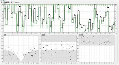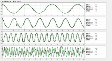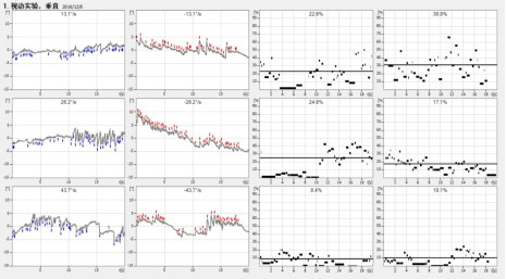
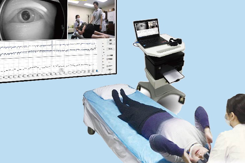
VertiPACS is the integrated software platform for all ZEHNIT vestibular products. It offers a complete arsenal of tests and procedures as well as a rich set of editing and documentation tools. Its basic features are:
• Patient database:
The easy-to-use database makes creating and accessing patient files simple.
• Network capabilities:
Examinations and assessment do not always happen in the same room. VertiPACS allows the same patient file to be accessed from different locations.
• Remote diagnosis capabilities:
With VertiPACS, you can send patient files via the Internet. This enables assessment by experts located anywhere in the world.
• Report generation:
Reports and summaries can be created as printouts or PDF files. Hospital logos can be edited and included, supporting your corporate identity.
• Editing and playback:
Some tests need a close look and sometimes even some editing to smooth out artifacts and make the result more clear-cut. Every test can be edited and played back.
• Device control:
VertiPACS controls all ZEHNIT hardware devices, including VertiGoggles, VertiPlatform, VertiChair and VertiSVV.
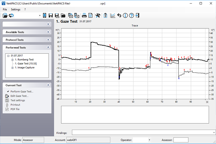
The VertiPACS test arsenal:
Documentation tools:
• History:
This section lets you document clinical findings like ear findings, vestibulo-spinal tests, neurological examinations, etc.
• Clinical findings:
This section permits the documentation of clinical findings like ear findings, vestibu-lospinal tests, neurological examinations, etc.
• Image capture:
Often, in the course of dizziness diagnostics, images are produced – photos of the eardrum taken during otoscopic examinations, pictures of the ocular fundus produced by ophthalmoscopes, or X-rays of the petrous part of the temporal bone. These images can be saved in the patient database and printed out as part of the report.
• Backseat Doctor: A small questionnaire that enables the VertiPACS KI to guess the most likely diagnosis. This test can be performed using VertiQuest, a smartphone app that is so simple that the patient can use it before even getting in touch with a doctor.
• Dizziness Handicap Inventory:
When this well-known questionnaire is used with VertiPACS, the program calculates the score and sub-scores and gives an instant result. This test can even be performed independently by a patient using VertiQuest.
• Gaze:
This test is performed to document gaze-dependent spontaneous nystagmus, auto-matically calculating its direction and strength.
• Supine roll/Position test:
This test is designed to document position-dependent nystagmus, which frequently presents in patients suffering from atypical BPPV or central nervous system disorders.
• Dix-Hallpike/Positioning test:
This test is performed to diagnose BPPV caused by displaced otoconia in the posterior semicircular canal.
• Caloric test:
This is the classic test of the labyrinth as devised by Barany. In addition to the analysis of spontaneous nystagmus, configurable diagrams display the Slow Phase Velocity (SPV). Vestibular paresis and directional preponderance are calculated and shown in tables.
• Custom test:
This feature allows users to freely configure their own goggles test.
• Head Impulse Test:
The Head Impulse Test (HIT) allows a side-dependent evaluation of the semicircular canals.
• SHIMP:
The new variant of the HIT offers a simpler assessment. It is known as the “covert saccades killer”.
•Optokinetic test:
Dizziness caused by a malfunctioning of the optokinetic system is indicative of a central nervous system disorder. This test allows an examination of the optokinetic system using different stimuli in a horizontal and vertical direction.
• Smooth Pursuit:
This test assesses the functioning of the brain’s pursuit center. A pathological outcome is indicative of degeneration or another type of central nervous system disorder.
• Predictive Pursuit:
This test allows an assessment of the patient’s ability to make predictive eye movements.
• Step-Ramp test:
Variant of the Smooth Pursuit test in which the initial saccades are suppressed by a short jump of the visual target.
• Gaze holding:
This examination is performed to detect gaze-dependent nystagmus. The patient’s gaze follows a visual target that moves incrementally from the center of the screen to the edge. If this triggers early nystagmus in the patient, it is a sign of a central disorder.
• Conventional Saccades:
This test allows the examiner to assess malfunctions of the saccade system.
• Advanced Saccades:
Gap Saccades, Overlap Saccades, Anti-Saccades, Predictive Saccades and Memory Saccades allow a detailed assessment of almost every part of the brain and so allow a kind of “brain mapping” with a sensitivity that can, in certain cases, be higher than an MRI.
• Romberg test:
This test allows a quantification of the Romberg test. It displays the result as a 2D Stabilogram and as a sway frequency diagram (Fourier analyzed).
• Balance training:
This set of exercises can be done by patients suffering from virtually any kind of balance disorder. Standing on the balance platform and using a monitor as a guide, the patient is trained to move his or her center of gravity in a targeted way. This feedback method increases the patient’s motivation and speeds compensation of vestibular loss.
• Subjective Visual Tests:
Combined with VertiSVV, these tests enable the user to perform the Subjective Visual Vertical as well as the Subjective Visual Horizontal test. In addition to the classic upright configuration, the SVV or SVH can be also performed in tilted positions to assess the utricle and saccule at the same time. The result is displayed in diagrams and tables.
• BPPV Chair test:
This is a rotary chair test for the diagnosis of BPPV. In addition to diagnostic uses, it allows the treatment of all kinds of BPPV. Predefined and user-defined procedures can be performed to determine and utilize the most effective treatments.
• Unilateral centrifugation (SVV):
This test is performed to assess the function of each utricle independently. First, the seated patient is rotated on the vertical axis until the chair reaches its final speed. Then the chair is moved a few centimeters to the side, just enough to bring one utricle into the center of the rotation. Now the function of the other utricle can be assessed, either by performing Subjective Visual Vertical tests (SVV) or by means of 3D oculography.
